 五度妙笔
五度妙笔 API商城
API商城
 数据库
数据库Glycobiology: How enzymes make the right choice during biosynthesis

Department of Biochemistry and Molecular Biology, University of Georgia, United States
Complex Carbohydrate Research Center, University of Georgia, United States
From prokaryotes to eukaryotes, cell surfaces are decorated with biopolymers that are essential for a wide range of processes, including cellular defense, environmental adaptation, intercellular communication, and tissue and organ structure (
Frantz et al., 2010
;
Whitfield et al., 2020
). These biopolymers are chemically complex, often consisting of thousands of repeating units. For example, hyaluronan – a biopolymer that plays vital structural roles in connective tissue, vascular tissue and cartilage in vertebrates – consists of more than 10,000 repeating units made of glucuronic acid (GlcA) and N-acetylglucosamine (GlcNAc) (
Cowman et al., 2015
;
Litwiniuk et al., 2016
).
Hyaluronan biosynthesis is a multistep process that involves alternately binding one of two substrates, UDP-GlcA and UDP-GlcNAc, to the catalytic site of an enzyme called hyaluronan synthase, which is embedded in the plasma membrane of the cell (
Figure 1
;
Górniak et al., 2026
;
Maloney et al., 2022
). This enzyme is also responsible for translocating the hyaluronan polymer through the plasma membrane, which allows it to extend into the extracellular space (
DeAngelis and Zimmer, 2023
). Hyaluronan synthases can be classified as processive (Class 1) enzymes or as distributive (Class 2) enzymes. Processive hyaluronan synthases can elongate either the non-reducing end of the polymer (Class 1-NR enzymes) or the reducing end (Class 1 R enzymes). On the other hand, distributive enzymes are only able to elongate the non-reducing end (
Blackburn et al., 2018
).
Figure 1
Download asset
Open asset
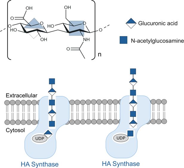
Structure, biosynthesis and translocation of the biopolymer hyaluronan.
Top: Hyaluronan is a linear biopolymer composed of repeating units made of glucuronic acid (GlcA) and N-acetylglucosamine (GlcNAc). Bottom: The enzyme hyaluronan synthase (pale blue) is embedded in the cell membrane (gray) and is responsible for the biosynthesis of hyaluronan, and also for its translocation across the cell membrane. Biosynthesis involves alternately adding GlcA and GlcNAc from UDP-linked substrates inside the cell to one end of the hyaluronan, while translocation involves pushing the hyaluronan chain into the extracellular space. Stephens et al. have helped shed light on this mechanism, ensuring that GlcA is always added when GlcNAc is at the end of the polymer chain (left), and vice versa (right).
Over the past decade, researchers have clarified many aspects of hyaluronan biosynthesis, but key mechanistic questions remain. In particular, how does a single active site discriminate between the two different substrates (UDP-GlcA and UDP-GlcNAc), and how is processivity – the ability to catalyze successive reactions without releasing the substrate – maintained? Now, in eLife, Zachery Stephens, Julia Karasinska and Jochen Zimmer of the University of Virginia School of Medicine report the results of experiments on hyaluronan biosynthesis in a virus called
Chlorella
that provide answers to some of these questions (
Stephens et al., 2026
;
Figure 1
).
The researchers used cryo-electron microscopy and enzyme assays to investigate the interaction between hyaluronan synthase and its substrates. Their results indicate that the enzyme distinguishes between UDP-GlcA and UDP-GlcNAc via a two-step recognition mechanism: the substrate first binds in a preliminary ‘proofreading’ position, where it is inspected, before being repositioned into the catalytic pocket so that GlcA or GlcNAc can be added to the growing biopolymer. Moreover, the efficient addition of GlcA requires the enzyme to be primed (that is, a sugar molecule must already be bound to the enzyme’s active site). The enzyme is usually primed by GlcNAc, which is a monosaccharide, but – somewhat surprisingly – Stephens et al. found that it can also be primed by two disaccharides – cellobiose and chitobiose. This reveals an unexpected flexibility in hyaluronan biosynthesis.
It is thought that class 1-NR hyaluronan synthases are structurally similar among different vertebrate species, indicating that the mechanism of hyaluronan biosynthesis is likely shared across vertebrates. However, there are notable differences between Class 1 R and Class 1-NR enzymes. For example, Class 1 R hyaluronan synthases from
Streptococci
bacteria have been shown to function as homodimers, with one member of the dimer positioning the acceptor chain, while the other binds the incoming donor substrate (
Blackburn et al., 2018
). This is in stark contrast to the way that Class 1-NR hyaluronan synthases function.
Future studies are needed to investigate Class 1 R hyaluronan synthases in more detail. The results of such studies, combined with the latest findings of Stephens et al., will lead to a deeper understanding of hyaluronan biosynthesis and could have applications in biomaterials production and hyaluronan-based therapies for wound healing and tissue repair.
References
Distinct reaction mechanisms for hyaluronan biosynthesis in different kingdoms of life
Glycobiology
28
:108–121.
PubMed
Google Scholar
The content and size of hyaluronan in biological fluids and tissues
Frontiers in Immunology
6
:261.
PubMed
Google Scholar
Hyaluronan synthases; mechanisms, myths, & mysteries of three types of unique bifunctional glycosyltransferases
Glycobiology
33
:1117–1127.
PubMed
Google Scholar
The extracellular matrix at a glance
Journal of Cell Science
123
:4195–4200.
PubMed
Google Scholar
Structural insights into translocation and tailored synthesis of hyaluronan
Nature Structural & Molecular Biology
32
:161–171.
PubMed
Google Scholar
Hyaluronic acid in inflammation and tissue regeneration
Wounds
28
:78–88.
PubMed
Google Scholar
Structure, substrate recognition and initiation of hyaluronan synthase
Nature
604
:195–201.
PubMed
Google Scholar
Insights into substrate binding and utilization by hyaluronan synthase
eLife
14
:RP109624.
PubMed
Google Scholar
Assembly of bacterial capsular polysaccharides and exopolysaccharides
Annual Review of Microbiology
74
:521–543.
PubMed
Google Scholar
Article and author information
Author details
Ana S Ramírez

"This ORCID iD identifies the author of this article:"
0000-0002-1619-0721
Publication history
Copyright
© 2026, Ramírez and Perez
This article is distributed under the terms of the
Creative Commons Attribution License
, which permits unrestricted use and redistribution provided that the original author and source are credited.
Metrics
0
citations
Views, downloads and citations are aggregated across all versions of this paper published by eLife.
Download links
A two-part list of links to download the article, or parts of the article, in various formats.
Downloads
(link to download the article as PDF)
Article PDF
Open citations
(links to open the citations from this article in various online reference manager services)
Mendeley
Cite this article
(links to download the citations from this article in formats compatible with various reference manager tools)
Glycobiology: How enzymes make the right choice during biosynthesis
eLife
15
:e111042.


